
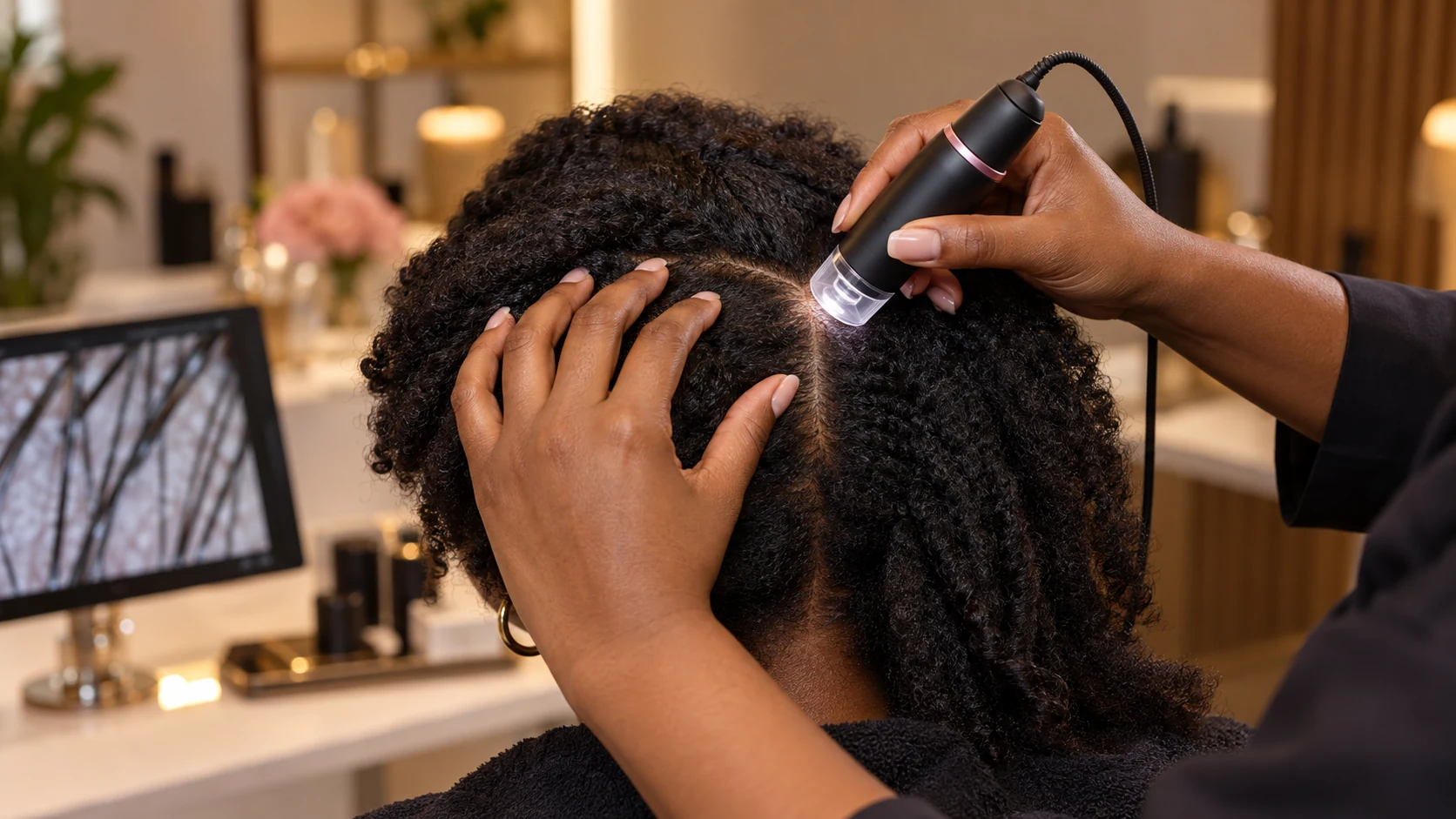
A scalp camera can make the hair and scalp easier to see, but magnification is not the same as a diagnosis. Trichoscopy is most useful when the images answer a focused question: Is density changing in a particular pattern? Are hairs breaking or becoming finer? Is there scale, redness, or another feature that needs medical attention?
A careful Hair & Scalp Analysis combines those images with your history, symptoms, styling practices, medications, and timeline. That context keeps a striking close-up from being mistaken for a complete explanation.
What Trichoscopy Is
Trichoscopy is a noninvasive method of examining the scalp and hair with magnification and directed light. A handheld scope or digital microscope captures close views of selected areas. Some systems display the image on a monitor and save photographs for comparison.
The examination may show hair-shaft diameter differences, the number of visible hairs in follicular openings, broken hairs, scale, redness, follicular debris, and the distribution of empty-looking openings. These observations can help organize the next step. They do not, by themselves, establish why a feature is present.
What Happens During a Scalp-Camera Appointment
The appointment should begin before the camera touches the scalp. The practitioner needs to know what changed, when it began, whether the change was sudden or gradual, and whether you have itching, burning, tenderness, bumps, heavy scale, shedding, or breakage.
Hair is then parted in more than one area. Images may be taken at the front, temples, center part, crown, sides, and back so one region can be compared with another. A localized concern may need close views of both the affected area and nearby hair that appears unchanged.
Good documentation records the location and uses consistent lighting, magnification, parting, and camera distance. Without that consistency, a later photograph may look different because the technique changed rather than because the hair changed.
What Magnified Images Can Help Show
Trichoscopy can help distinguish a primarily scalp-surface concern from a change in the hair shafts or distribution. It can make short broken pieces easier to separate from full-length shedding and can reveal whether hairs within one area vary noticeably in diameter.
It can also support a more precise referral. For example, a smooth localized patch, a widening part, a tender crown with scale, and breakage around a high-tension style are different patterns. Clear images and a reliable timeline give a dermatologist or other clinician better information than the general statement that “my hair is falling out.”
Magnification is particularly useful for baseline documentation. If care is changed, later images can be compared with the same scalp locations rather than relying only on memory or day-to-day mirror checks.
What a Scalp Camera Cannot Confirm
A camera cannot determine iron status, thyroid function, hormone levels, nutritional intake, medication effects, autoimmune activity, or the internal phase of every follicle. It cannot replace laboratory testing when a clinician considers testing appropriate.
It also cannot reliably rule out a scarring process simply because the skin looks calm in one photograph. Some hair-loss conditions require medical examination, dermoscopy by a dermatologist, laboratory work, fungal testing, or a scalp biopsy. A responsible analysis explains those limits instead of assigning a diagnosis from one enlarged image.
Analysis, Assessment, and Medical Diagnosis
A trichology-focused analysis can document the pattern, review cosmetic and styling factors, identify visible concerns, and help determine whether medical referral should come first. It may also support a nonprescription scalp-care or hair-care plan within scope.
A dermatologist or other qualified medical clinician diagnoses disease, orders tests, prescribes medication, and performs procedures such as biopsy. These roles can complement each other. The safest path is not to force every concern into one provider's scope.
Who May Benefit From Trichoscopy
Magnified documentation can be useful when a part appears wider, the crown looks different in photographs, shedding has increased, breakage and shedding are difficult to separate, or scalp symptoms keep returning. It may also help before an extension or protective-style plan when density appears uneven.
Someone with stable hair who wants a baseline may benefit, but more imaging is not automatically better. The examination should have a purpose and a plan for what happens if a concern is seen.
When Medical Care Should Come First
Seek timely medical evaluation for a rapidly expanding bald patch, pain or burning, pus, bleeding, open sores, heavy crusting, loss of follicular openings, a shiny area, eyebrow or body-hair loss, or hair change with systemic symptoms. Children with a scaly patch or broken hairs may need prompt evaluation for a contagious fungal infection.
Sudden severe shedding, suspected medication effects, pregnancy-related concerns, or symptoms suggesting anemia, thyroid disease, or another medical condition also belong with a licensed healthcare professional. A scalp-camera appointment should not delay necessary care.
How to Prepare for Useful Images
Follow the preparation instructions from the practice. Unless told otherwise, avoid applying fresh hair fibers, colored scalp sprays, heavy oils, glitter, or thick styling product immediately before the visit because they can cover the scalp surface and alter what the camera records.
Bring a medication and supplement list, relevant diagnoses, recent laboratory results if you already have them, and a timeline of illness, surgery, childbirth, major stress, weight change, chemical services, or tight styling. Photos from before the change can help establish your baseline.
Questions to Ask Before Booking
Ask who performs the analysis, what training they have, whether images are stored, how privacy is handled, what the service can and cannot determine, and when referral is recommended. Ask whether the appointment includes written next steps and whether follow-up images use the same locations and settings.
Be cautious with claims that a camera can identify every deficiency, diagnose disease instantly, or guarantee regrowth. Technology improves observation; it does not remove the need for clinical judgment and appropriate scope.
Why Comparison Areas Matter
A close-up of the area that worries you is only half of a useful examination. The practitioner should also look at one or more comparison areas. Hair diameter and the number of hairs emerging together naturally vary across the scalp, and normal features can appear unusual when they are enlarged without a reference.
For a widening center part, comparison images may come from the sides or back. For a temple concern, the opposite temple and a stronger interior area can provide context. When breakage is suspected, the camera may follow several strands from the scalp outward to look for weathering, splitting, or abrupt ends.
Comparison does not create a diagnosis, but it reduces the chance that one dramatic image drives the entire conclusion. It also creates a more reliable baseline for follow-up. If treatment or routine changes are made, progress should be judged with the same locations and technique, not by selecting whichever photograph looks most favorable.
Using the Findings to Choose the Next Step
The most useful outcome is a clearer decision. That might be a dermatologist referral, a conversation with your primary-care clinician, a change in styling tension, a gentler cleansing plan, documented monitoring, or a separate consultation about extensions after stability is established.
GlamorChiQ offers virtual and in-person Hair & Scalp Analysis. In-person appointments can include direct scalp-camera imaging, while virtual appointments rely on history and client-supplied photographs or video. Neither format replaces medical diagnosis when symptoms or findings call for it.
Turn close-up images into a clear next step
Choose the analysis format that fits your location and concern. Your history, pattern, symptoms, and images are reviewed together.
Choose Your AnalysisFrequently Asked Questions
Trichoscopy is a noninvasive, magnified examination of the scalp and hair. A handheld digital scope can document features that are difficult to judge with the unaided eye, but the images must be interpreted in context.
Trichoscopy can support pattern recognition and documentation, but a camera image alone does not replace a medical history, physical examination, laboratory testing, biopsy, or diagnosis by a licensed medical clinician when those are needed.
The imaging itself is generally painless. Hair is parted and the camera is placed close to or lightly against selected areas of the scalp. Tell the practitioner if an area is tender or inflamed.
Follow the instructions provided for your appointment. In general, arriving with the scalp visible and without fresh heavy oils, fibers, sprays, or color products can make observation easier, but do not change prescribed scalp treatment unless your clinician directs you.
The findings are reviewed alongside your history, symptoms, routine, styling practices, medications, and pattern of change. The next step may be routine changes, progress photography, referral to a dermatologist or other clinician, or a follow-up plan within the practitioner’s scope.